
pain at top of calf behind knee treatment
Pain at the Top of the Calf Behind the Knee: How to Decide What to Do Next

Route the symptom before naming a cause
Pain where the back of your knee meets the upper calf needs a care decision before it needs a label. The same general area can hurt after physical activity, with an orthopedic problem, or with a blood-vessel problem, and the location alone cannot establish which explanation applies to you.
Use this order:
- Check for symptoms that change the timeline. Signs or symptoms of deep-vein thrombosis or pulmonary embolism require immediate care. The CDC advises immediate care when symptoms of deep-vein thrombosis or pulmonary embolism occur.
- Separate injury-related danger signs. Severe pain, swelling, instability, or an inability to bear weight after an acute injury warrants urgent medical evaluation. Fever with swelling, redness, and limited motion also needs urgent attention.
- If those warning signs are absent, use the symptom pattern as context rather than a diagnosis. Activity-related pain may be muscular, but sudden calf pain without a clear cause can signal a serious blood-vessel problem.
- Arrange an evaluation when pain persists, worsens, or limits movement. A clinical history and examination can help determine whether monitoring, testing, or treatment discussion makes sense.
The goal is not to decide at home whether your pain is a tendon, muscle, joint, nerve-related, or blood-vessel problem. The goal is to choose the appropriate level and timing of care. That distinction matters whether you live in Ohio, Chicago, or elsewhere in the United States: a possible treatment is not the first decision when the symptom could need urgent assessment.
First check for symptoms that change the timeline
Warning signs involving breathing, consciousness, or possible blood clots require prompt medical assessment rather than routine treatment planning. NICE guidance identifies chest pain, fainting or near-fainting, rapid breathing, and features of deep-vein thrombosis as warning features in the assessment of possible pulmonary embolism. The CDC also advises immediate care when symptoms of deep-vein thrombosis or pulmonary embolism occur.
Seek immediate medical attention if pain near the knee or calf occurs with:
- Chest pain.
- Fainting or near-fainting.
- Rapid breathing.
- Signs or symptoms that suggest a blood clot.
Do not use the absence of a sports injury to dismiss this route. A sudden episode without a clear cause deserves more caution than soreness that began after a specific physical demand, although neither pattern diagnoses the problem. If these symptoms are present, do not wait for a regenerative consultation or a routine orthopedic appointment to decide what is happening.
NICE guidance recommends immediate hospital admission for suspected pulmonary embolism when there are signs of hemodynamic instability. For other suspected cases, clinicians use a clinical assessment and testing pathway rather than relying on location alone. You do not need to calculate a clinical score yourself; the practical boundary is to seek prompt medical assessment when these warning features appear.
This warning-sign screen is separate from deciding whether a musculoskeletal consultation might help. A consultation can be considered only after symptoms requiring immediate attention have been addressed. If you are unsure whether a symptom represents a blood-clot warning sign, treat uncertainty as a reason to seek prompt medical guidance rather than testing yourself by walking farther.
Treat injury-related danger signs as a separate route
Injury-related pain needs urgent evaluation when the injury is accompanied by severe pain, swelling, instability, or an inability to bear weight. These findings can indicate a problem that should be assessed promptly instead of watched through continued activity.
Fever with swelling, redness, and limited motion is another urgent pattern because it can signal a joint infection. That combination does not become routine simply because the pain is located behind the knee rather than in the front of the joint.
Use the event that preceded the pain as part of the decision:
- After an acute injury: A fall, twist, collision, sudden change of direction, or other clear event combined with severe pain, swelling, instability, or inability to bear weight calls for urgent medical evaluation.
- With possible infection signs: Fever with swelling, redness, and limited motion needs urgent medical attention.
- Without those findings: Continue to the symptom-pattern questions below, but do not treat a lack of obvious injury as proof that monitoring is appropriate.
A clear injury can help a clinician understand what to examine, but it does not tell you at home whether the muscle, tendon, joint, or another structure is involved. The correct action in this route is evaluation, not an attempt to identify the injured structure by repeatedly loading it.
Use onset and activity as context, not a diagnosis
The timing and trigger of pain behind the knee provide useful context, but they do not establish a diagnosis. Pain in this region can also occur with orthopedic conditions such as a torn muscle, damaged tendon, or inflamed joint.
Record the sequence rather than assigning a cause:
- Note whether the pain began during activity, after activity, or without a clear trigger.
- Describe whether it started suddenly or built over time.
- Record the exact location, such as the crease behind the knee, upper calf, or a broader area.
- Note whether swelling, redness, instability, weakness, numbness, or limited motion appeared with it.
- Record what happens when you walk, climb stairs, sit, stand, or rest, without repeatedly provoking the symptom to test it.
Activity-related onset is information to bring to an examination. It cannot rule out a more serious problem, and a sudden unexplained onset should not be treated as ordinary post-exercise soreness.
A useful record separates observation from interpretation. “Pain began after I increased walking” is information you can bring to an appointment. “I strained a tendon” is a conclusion that requires clinical assessment. Keeping those statements separate helps you avoid choosing treatment before the problem has been identified.
Keep the possible categories broad
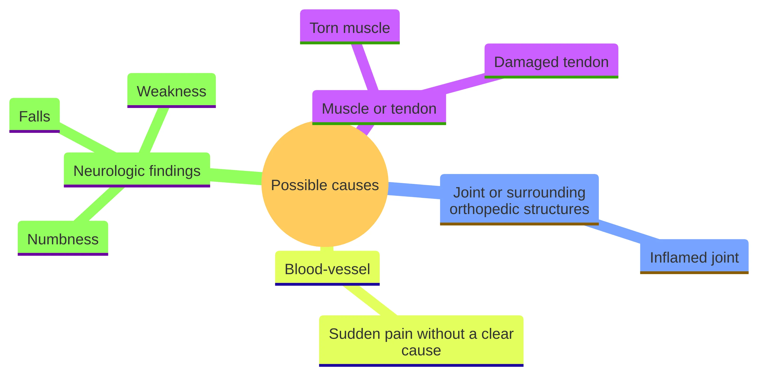
Possible causes of pain behind the knee and at the top of the calf belong to several broad categories, and your symptoms may fit more than one question until you are examined. Orthopedic causes can include a torn muscle, damaged tendon, or inflamed joint.
A useful category check looks like this:
- Muscle or tendon: A torn muscle or damaged tendon remains a possibility that requires examination rather than a conclusion based on location.
- Joint or surrounding orthopedic structures: An inflamed joint can produce pain in the area behind the knee. Fever, swelling, redness, and limited motion make this an urgent assessment question.
- Blood-vessel: Chest pain, fainting or near-fainting, rapid breathing, or other signs of a blood clot require prompt medical assessment.
- Neurologic findings: Weakness, numbness, falls, or other neurologic symptoms are details to report because they affect the clinical assessment. They do not identify the source by themselves.
This broad approach prevents a common mistake: treating a familiar symptom pattern as proof that the pain is muscular. It also prevents the opposite mistake of assuming that every symptom behind the knee has the same urgent explanation. Keep the category open until the history and examination narrow it.
The distinction is practical. The same complaint may lead to different questions about weight-bearing, circulation, strength, motion, sensation, or further testing. A location label cannot answer those questions, and a treatment that might be discussed for one musculoskeletal condition should not be assumed to apply to unexplained calf pain.
Do not make walking the test
Walking is a function to observe, not a diagnostic test. Pain during walking or running is generally muscular according to the cited clinical guidance, but that general pattern does not tell you whether your pain comes from a muscle, tendon, joint, or blood vessel.
Use ordinary movement only to describe what happens:
- If walking brings on pain, record when it starts and whether it changes your stride.
- If walking makes the pain worse, do not repeatedly walk farther, faster, or uphill to prove what is wrong.
- If you cannot bear weight after an acute injury, treat that finding as a reason for urgent medical evaluation.
- If walking is possible but pain continues, worsens, or limits normal movement, arrange an evaluation instead of using continued walking to settle the cause.
This section does not prescribe rest, exercise, or another individualized activity plan. Until a clinician assesses the pattern, do not use walking or repeated loading to test the cause; record what ordinary movement does and arrange evaluation if pain persists, worsens, or limits function. Until that assessment, avoid turning a painful movement into a repeated experiment or assuming that tolerating a short walk means the condition is minor.
You can still describe function without challenging the symptom. Note whether you can walk normally, whether you are shortening your stride, and whether stairs or standing change the pain. That information helps an appointment begin with a useful functional picture instead of a self-administered stress test. For general activity, do not repeatedly walk farther, faster, or uphill to test the cause; use ordinary movement only to note symptoms and arrange evaluation when pain persists, worsens, or limits function.
Let persistence and function set the evaluation boundary
Persistent or function-limiting pain deserves an evaluation even when no emergency warning sign is present. The effect on sleep, movement, walking, and daily tasks gives a clinician information about the problem and helps determine whether further testing is needed.
Arrange a medical evaluation when:
- Pain continues instead of steadily settling.
- Pain is getting worse or returning with ordinary activity.
- Pain changes how you walk, stand, climb stairs, or sleep.
- Weakness, numbness, falls, or other neurologic symptoms accompany the pain.
- You cannot explain the onset or the pattern keeps changing.
- The symptoms interfere with work, exercise, caregiving, or other normal responsibilities.
Do not wait for a fixed number of days if urgent symptoms, severe pain, or inability to bear weight appears. Those findings change the route immediately. If the problem is persistent but stable, a routine clinical evaluation can clarify whether the next step is observation, testing, or a treatment discussion.
The boundary is functional as well as chronological. Pain that keeps you from doing normal activities gives you a reason to stop indefinite self-monitoring, even if the pain has not reached an emergency level. You do not need to prove that the pain is severe enough by continuing the activity that aggravates it.
Bring the change over time to the appointment: whether the area is becoming more painful, whether the affected area is expanding, whether movement is becoming harder, and whether new symptoms have appeared. Those observations are more useful than trying to convert persistence into a diagnosis.
Bring a symptom timeline that can be examined
A symptom timeline gives your clinician a clearer starting point than a treatment request based only on location. Evaluation of knee-area pain usually considers when the pain began, where it is, what it feels like, associated mechanical or systemic symptoms, swelling, any precipitating trauma, and relevant medical or surgical history.
Before an appointment, write down:
- Onset: The date or approximate period when symptoms began, and whether the onset was sudden or gradual.
- Location: The most painful point and whether the discomfort spreads into the calf, thigh, or joint area.
- Quality: Aching, sharp, pulling, burning, pressure, or another description that matches your experience.
- Triggers: Walking, running, stairs, bending, standing, sitting, or rest.
- Function: Whether you can bear weight, walk normally, move the knee, sleep, and complete ordinary tasks.
- Associated findings: Swelling, redness, fever, instability, weakness, numbness, falls, chest pain, fainting, rapid breathing, or other symptoms.
- Context: Recent injury, change in activity, prior surgery, relevant medical history, and treatments you have already tried.
A first pain-management visit may also cover what changes the symptoms, how they affect sleep and movement, previous treatments, and whether weakness or numbness is present. Bring imaging reports or prior records if you have them, but do not assume that an old image explains a new symptom.
Also write down what you want the visit to answer. You might ask whether the pattern calls for urgent assessment, whether the examination suggests a musculoskeletal problem, whether further testing is needed, and which options remain if regenerative treatment is not appropriate. A focused list keeps the appointment centered on the decision you need to make.
To make the timeline more useful, separate what you observed from what you want answered. For example, list the trigger and change in function as observations, then ask whether the pattern calls for examination, testing, or another pathway. Keep a short record of what you tried and what happened afterward; this gives the clinician context without turning the response into proof of a diagnosis. If you have prior imaging, note the date, body part, and reason it was obtained. The clinician can then judge whether it relates to the current symptoms rather than assuming that any prior result is decisive.
You can also organize the visit around three decisions: whether anything needs prompt attention, what the examination is intended to clarify, and what options remain after the assessment. A clear answer may be that regenerative treatment is not appropriate. That answer is clinically useful because it prevents an undiagnosed or unsuitable problem from being treated as though the desired option were already selected.
If symptoms change before the appointment, update the timeline rather than relying on your first description. New chest symptoms, fainting, rapid breathing, or other possible blood-clot signs move the situation out of routine appointment planning and into prompt medical assessment.
Understand what clinical assessment is trying to distinguish
Clinical assessment of pain behind the knee and upper calf is designed to connect your symptom pattern with the structures and risks that need attention. A systematic knee examination may include looking at and feeling the area, testing motion and strength, checking nerves and circulation, and performing specific maneuvers.
Your history and examination work together. The clinician may ask whether the pain followed trauma, whether swelling or systemic symptoms are present, and how the problem affects movement. The examination can then compare motion, strength, circulation, sensation, and stability rather than relying on the painful location alone.
A first pain-management visit may also include a review of imaging, a physical examination, and a discussion about whether further testing is needed. Be ready to describe weakness, numbness, falls, and other neurologic symptoms even if the pain itself feels muscular. Those details can change what the clinician needs to examine.
An assessment may therefore answer several different questions:
- Is there a warning pattern that requires prompt care?
- Did an acute injury create a problem with weight-bearing, stability, or motion?
- Does the pattern fit an orthopedic question that needs further evaluation?
- Do circulation or neurologic findings require a different care pathway?
- Is testing needed before any treatment conversation?
The purpose is not to force your symptoms into a named condition. It is to identify the next clinical question safely. The examination may support continued observation, indicate that testing is needed, or show that routine musculoskeletal treatment planning is not the right route.
That is why a request for a particular injection or regenerative therapy should remain secondary to the evaluation. The treatment discussion depends on what the history and examination establish, not merely on the place where you feel pain.
Each part of the assessment contributes a different kind of information. Looking at and feeling the area can document visible or local changes; motion and strength testing can show how function is affected; nerve and circulation checks address findings that may point beyond a simple local muscle complaint; and specific maneuvers can help the clinician compare possible orthopedic explanations. These components do not provide a home diagnostic formula. Their value comes from being interpreted together with onset, associated symptoms, injury history, and functional change.
That combined view also explains why the same symptom can produce different next steps. If the history emphasizes an acute event and weight-bearing difficulty, the immediate question is different from a persistent pattern without a clear trigger. If weakness, numbness, falls, or circulation-related concerns are reported, the clinician may need to consider a different pathway than a treatment discussion focused only on a sore area. If the assessment supports a musculoskeletal problem, the next discussion can be about whether testing or a non-surgical option is appropriate; if it does not, forcing a regenerative plan would not answer the underlying question.
Before accepting a proposed plan, ask what finding led to it and what uncertainty remains. You can ask which part of the history or examination supports the working explanation, whether a test is needed to resolve an important uncertainty, and what alternative route would be used if the findings do not fit. These questions do not require you to name the condition yourself. They help you understand how the evaluation leads to the proposed next step.
Match testing to the clinical question
Testing for pain behind the knee should follow the clinical question rather than the desire for an immediate image. For knee pain, X-rays are generally reserved for chronic pain lasting more than six weeks or for certain acute injuries that meet evidence-based criteria. The decision depends on the history and examination, not on the fact that the pain is uncomfortable.
Possible testing routes are not interchangeable:
- Orthopedic questions: The clinician may decide whether an examination is enough initially or whether imaging is justified by chronic symptoms or an acute injury that meets specific criteria.
- Blood-clot or pulmonary questions: Suspected pulmonary embolism requires clinical evaluation and testing. – Unclear or changing patterns: The next test depends on what the history and examination show. A test selected for a joint or bone question does not automatically answer a blood-vessel question.
Do not treat a normal or absent image as a complete answer without understanding what the test was intended to evaluate. Ask what question the test is meant to answer, what the result would change, and whether another care pathway is needed if the symptom pattern does not fit the initial assumption.
The timing also matters. An X-ray decision for persistent knee pain is not the same as the urgent evaluation pathway used when clinicians suspect a pulmonary embolism. These routes address different clinical questions, so you should not use the general knee-imaging boundary to delay care for warning symptoms involving breathing, consciousness, or possible blood clots.
Testing should be understood as a way to answer a defined clinical question, not as a measure of how concerning pain feels. An X-ray may be appropriate under the chronic-pain or qualifying-acute-injury criteria described above, but that does not determine how possible clot or pulmonary symptoms should be assessed. Conversely, an urgent assessment for those symptoms is not replaced by a joint-focused image or delayed until a musculoskeletal time threshold is reached.
Before a test, ask what structure or risk the clinician is evaluating and what information the result may add. If imaging is proposed, ask how its result would change the next step. If the history raises a circulation or neurologic question, ask how that question will be examined rather than assuming that an X-ray answers it. If an older result is reviewed, explain when it was obtained and whether the current symptoms are different.
A decision not to test immediately can still be part of a clinical plan, but it should be connected to the history and examination and to the change that would warrant reassessment. Persistent, worsening, or function-limiting pain should not disappear into an undefined waiting period. New warning symptoms change the route immediately.
The NICE pathway discussed in the supporting guidance is United Kingdom guidance, not a universal U.S. protocol. In the United States, clinicians use applicable local protocols and clinical judgment. This distinction does not change the immediate safety point: concerning symptoms should receive prompt medical assessment rather than a delayed joint-focused workup. It does change how to interpret the testing details, because the applicable process may depend on the clinical setting and local practice.
When a test is proposed, ask what clinical question prompted it, which structure or risk it evaluates, what a result would change, and what follow-up is planned if the result is normal, abnormal, or inconclusive. These questions are especially useful when symptoms cross categories, because a test aimed at an orthopedic structure may not answer a vascular or neurologic question. They also make clear why evaluation comes before selecting a treatment.
Discuss treatment only after suitability is established
Treatment discussion belongs after urgent causes have been addressed and the clinical assessment identifies a musculoskeletal problem that can reasonably be considered for treatment. A treatment name cannot substitute for knowing what structure is involved, whether testing is needed, and whether the option fits your condition.
Regenerative options also require an evidence-aware conversation. A systematic review of 23 studies reported early pain relief and functional gains with platelet-rich plasma and sustained effects with stem-cell therapies, but it found substantial variation in protocols and outcomes. Those findings describe research across musculoskeletal injuries; they do not establish that a regenerative treatment will help your undiagnosed pain, and they do not guarantee relief or avoidance of surgery.
A useful treatment conversation should address:
- What condition is being treated and how the assessment supports that conclusion.
- Whether the proposed option is directed at the suspected tissue or problem.
- What is known, uncertain, or variable about the option.
- What alternatives remain available, including a decision not to proceed.
- What timing and cost would apply if the option is clinically appropriate.
- What would happen if the assessment suggests that regenerative care is unlikely to help.
REGEN Orthopedic Medicine provides patient-centered, honest assessments and guides people toward alternative treatment when regenerative care is unlikely to help a joint. If surgery is deemed necessary, we inform the patient. Those boundaries keep a consultation focused on suitability rather than a promised result.
The practical question is not whether a regenerative treatment sounds appropriate from a description of symptoms. It is whether the clinical assessment identifies a condition, tissue, and treatment pathway for which that discussion is relevant. If the evaluation points elsewhere, follow that clinical direction rather than forcing the symptom into a regenerative-treatment category.
Suitability also includes the boundaries of the evidence and the purpose of the visit. The review cited here found variation in protocols and outcomes, so the name of a modality does not by itself describe the protocol, target, timing, expected course, or likely fit. Those details must be discussed in relation to the assessed condition, not inferred from a general claim about regenerative care.
A patient can make the conversation more concrete by asking: What finding makes this option relevant? What other options should I consider? What would make you advise against it? How will timing and cost be explained? What is the plan if the assessment points to surgery or another form of care?
The goal of this conversation is informed choice, not treatment selection by symptom location. The assessment may support a tailored non-surgical discussion, but it may instead support additional evaluation or an alternative recommendation. Keeping those possibilities open is consistent with an honest assessment and with the stated boundary that surgery should be discussed when it is deemed necessary.
What a REGEN Orthopedic Medicine consultation can cover
A consultation with REGEN Orthopedic Medicine can cover evaluation and non-surgical options for knee, hip, or shoulder pain when a musculoskeletal cause is relevant.
Our Ohio practice uses the body’s own biology, including stem cells, exosomes, and peptides, to treat knee, hip, and shoulder conditions without surgery. That description applies to the conditions and service scope stated by REGEN Orthopedic Medicine; it does not mean that every cause of pain near the calf or behind the knee is appropriate for regenerative care.
A consultation may help organize the next discussion around:
- The onset, location, quality, triggers, and functional effect of your symptoms.
- Whether the pattern appears relevant to a musculoskeletal evaluation or needs another care pathway first.
- Whether further testing or records should be considered before treatment planning.
- Whether a non-surgical regenerative option is clinically appropriate for the condition being assessed.
- Options, timing, and cost details if regenerative treatment is appropriate.
REGEN Orthopedic Medicine tailors each treatment option to your specific needs. If an evaluation points to peroneal tendonitis, our tailored treatment plans for that condition are led by fellowship-trained orthopedic surgeons. That example does not diagnose peroneal tendonitis from pain location alone.
The practical boundary is straightforward: a consultation can help determine whether the symptom belongs in a musculoskeletal treatment conversation, but it cannot make urgent warning signs safe to ignore or guarantee that a regenerative option will fit. If you have already addressed urgent symptoms and your pain persists or limits function, an evaluation can be a reasonable way to clarify the next question.
Free consultations are available for knee, hip, or shoulder pain at Ohio and Chicago locations, and no referral is needed. Most consultations begin as a telehealth call, so a patient can start without traveling to a clinic. These access details make it easier to ask an initial evaluation question, but they do not change the triage boundary: possible clot or pulmonary symptoms, severe injury-related findings, or possible infection signs need prompt medical attention rather than a routine consultation.
If a consultation proceeds, ask the team to explain what the assessment supports, whether further testing is needed, and whether the issue falls within the conditions for which non-surgical regenerative options are considered. If it does, REGEN Orthopedic Medicine provides options, timing, and cost details; if it does not, the consultation should remain an evaluation rather than a commitment to a procedure.
Choose the appropriate request, not a promised treatment
The next step for pain behind the knee and upper calf depends on the symptom pattern: seek immediate care for chest pain, fainting or near-fainting, rapid breathing, or signs of a blood clot; seek urgent evaluation for severe pain, swelling, instability, inability to bear weight after injury, or fever with swelling, redness, and limited motion; and arrange a clinical evaluation when pain persists, worsens, or limits function without those warning signs.
A qualified healthcare professional must determine what condition you have and whether any treatment is suitable for you. If urgent warning signs have been addressed and a musculoskeletal cause may be relevant, a consultation can help you review the symptom timeline, understand what assessment or testing may be needed, and discuss appropriate options, timing, and cost when regenerative treatment is clinically appropriate.
Questions to ask before requesting an evaluation
What should I do if I experience chest pain or fainting along with calf pain?
Seek immediate medical attention if you experience chest pain, fainting, rapid breathing, or signs of a blood clot along with calf pain, as these symptoms require prompt assessment.
When is it necessary to seek urgent medical evaluation for calf pain?
Urgent evaluation is necessary if you experience severe pain, swelling, instability, or inability to bear weight after an injury, or if you have fever with swelling, redness, and limited motion.
What information should I prepare for a healthcare visit regarding my calf pain?
Prepare a timeline of your symptoms, including when they began, their location, quality, triggers, and any associated symptoms like swelling or numbness. This information will help your clinician assess your condition.
What types of treatments does REGEN Orthopedic Medicine offer for joint pain?
REGEN Orthopedic Medicine offers non-surgical regenerative treatments such as stem cell therapy, exosome therapy, and peptide therapy for joint pain in the knee, hip, and shoulder.
Are consultations available at REGEN Orthopedic Medicine without a referral?
Yes, REGEN Orthopedic Medicine offers free consultations for knee, hip, or shoulder pain without the need for a referral.
What should I do if my calf pain persists or worsens?
If your calf pain persists, worsens, or limits your movement, arrange for a medical evaluation to determine the appropriate next steps.
Free consultation
Find out whether regenerative treatment is right for you.
Most consultations begin as a telehealth call with a physician specialist, so you can start without travelling to a clinic. No referral needed.